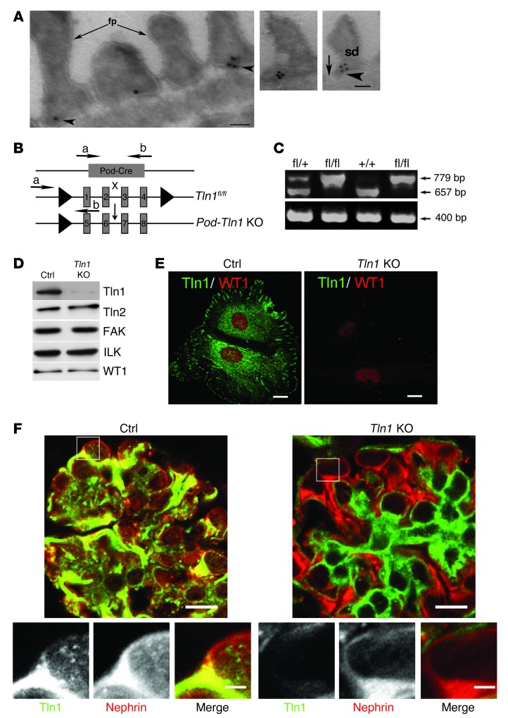
Figure 1. Generation of podocyte-specific Tln1-KO mice.
(A) Immunogold transmission EM staining demonstrates talin1 (arrowheads) localizes to the base of the foot process (fp) and adjacent to the slit diaphragm (SD) (arrows) of podocytes from WT mouse kidneys. Scale bars: 100 nm. (B) Schematic demonstrating breeding of the Podocin-Cre mice with Tln1fl/fl mice to generate podocyte-specific Tln1-KO mice (Pod-Tln1–KO). Forward and reverse primers are denoted as a and b, respectively, for Cre and Tln1. (C) Identification of Tln1 and Podocin-Cre by tail genotyping (age P7). (D and E) Talin1 expression in purified control (Ctrl) podocytes and lack of talin1 expression in podocytes harvested from Pod-Tln1–KO mice (age P7), as detected by Western blotting. (D) and immunofluorescence (E). Note that Ctrl podocytes express both talin1 and talin2. Podocytes were plated on collagen type I–coated glass coverslips and stained for WT1 (red) and for talin1 using an isoform-specific talin1 monoclonal Ab. Scale bars: 10 μm. (F) Double-immunofluorescence detection of nephrin (red) and talin1 (green) on kidney sections of the indicated genotypes (age P14). Scale bars: 10 μm (upper panels) 2 μm (lower panels).

