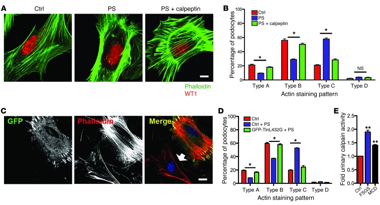
Figure 9. Increased calpain activity results in actin-cytoskeletal disorganization.
(A) Representative image of podocyte phalloidin staining following stimulation with protamine sulfate with or without calpeptin. Scale bar: 10 μm. (B) Quantification of the phalloidin staining pattern. n = 4 experiments. *P < 0.05, control or protamine sulfate plus calpeptin vs. protamine sulfate alone. (C) Representative image of primary podocytes expressing the calpain-resistant talin1 mutant (GFP-Talin L432G) stained with phalloidin following stimulation with protamine sulfate. Note loss of stress fiber in neighboring untransfected WT control (arrow). Scale bar: 10 μm.(D) Quantification of phalloidin staining pattern of C. n = 3 experiments. (E) Increased urinary calpain activity is observed in patients with biopsy-proven FSGS and MCD when compared with healthy controls. n = 17 patients with FSGS, 15 patients with MCD, and 6 healthy controls. *P < 0.05; **P < 0.01.

