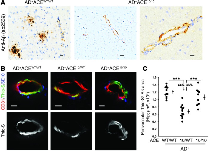
Figure 2. Perivascular Aβ deposition in AD+ACE10/WT and AD+ACE10/10 mice.
(A) Micrographs of paraffin sections from AD+ACEWT/WT and AD+ACE10/10 cerebral cortex (8.5-month-old mice) labeled for human Aβ using rabbit polyclonal antibody AB2539 with standard peroxidase-based techniques. AD+ACE10/10 mice showed perivascular deposition of Aβ. (B) Representative images of fluorescence-based immunohistochemistry and Thio-S staining. PFA-fixed hippocampal sections of 7- to 8-month-old mice were colabeled with Thio-S (green), anti-CD31 (red), an EC marker, and 6E10 mAb (blue). The Thio-S signal is also shown in gray scale (lower panels). Scale bars: 10 μm. (C) Quantitation of perivascular Thio-S+ area in blood vessels from the hippocampus was performed using serial coronal sections. Data for individual mice as well as for group means and SEM are indicated, along with the percentages of reduction in the mean plaque area. Perivascular Aβ deposition was significantly reduced in AD+ACE10/WT mice compared with that in AD+ACEWT/WT mice or AD+ACE10/10 mice. There was no statistical difference between the AD+ACE10/10 and AD+ACEWT/WT mice. n = 10–12 mice per group. ***P < 0.0001.

