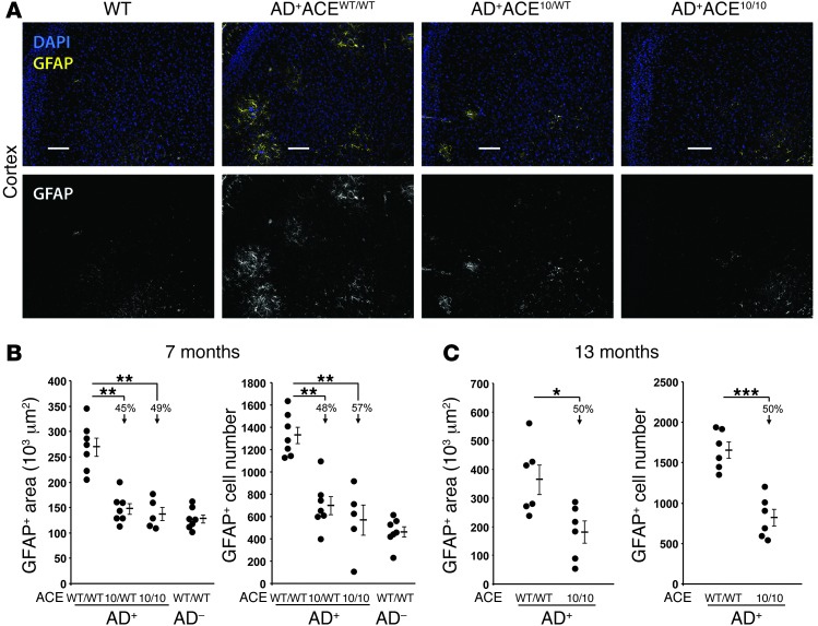
Figure 3. Reduced astrogliosis in AD+ACE10/10 and AD+ACE10/WT mice.
Coronal brain sections from 7- to- 8-month-old mice were stained with a polyclonal antibody against astrocyte-specific glial fibrillary acidic protein (GFAP; yellow). Sections were counterstained for nuclei with DAPI (blue). (A) Fluorescent micrographs of coronal brain sections show reduced staining for cortical GFAP in AD+ mice heterozygous or homozygous for the ACE 10 allele (AD+ACE10/WT and AD+ACE10/10). For simplicity of viewing, the bottom portion of panel A shows the GFAP channel as gray scale. Scale bars: 100 μm. (B) Quantitative analysis was performed to measure the GFAP+ area and cell numbers in 7- to 8-month-old mice. Data for individual mice are shown as well as for the group means and SEM. Arrows indicate the percentage of reduction in the group means as compared with that in AD+ACEWT/WT mice. Mice heterozygous or homozygous for the ACE 10 allele had significantly reduced astrogliosis. (C) A similar analysis of astrogliosis was performed on 13-month-old mice. Even at this age, AD+ACE10/10 mice had significantly fewer GFAP+ reactive astrocytes than did AD+ACEWT/WT mice. n = 5–7 mice per group. *P < 0.05; **P < 0.001; ***P < 0.0001.

