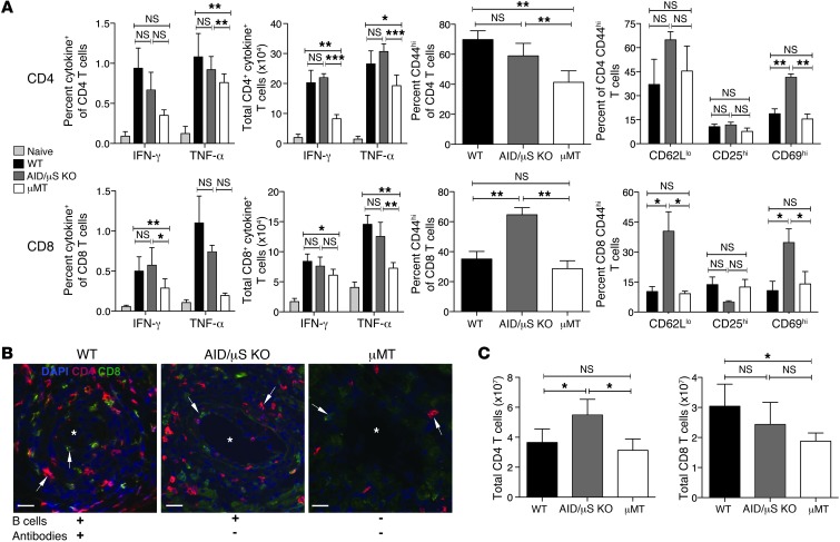
Figure 2. Alloreactive T cell responses are diminished in the absence of B cells.
(A) Spleen, LN, and BM cells were harvested from WT, AID/μS KO and μMT recipients of BALB/c hearts treated with costimulation blockade for assessment of T cell activation and cytokine production (at allograft harvest for CAV, 100–110 days after transplantation). Harvested cells were stimulated with BALB/c splenocytes for 6 hours and IFN-γ+ and TNF-α+ cells within the H-2d negative recipient T cells were assessed by intracellular cytokine staining. Percentage and total numbers of IFN-γ+ and TNF-α+, CD4+ and CD8+ T cells in allograft recipients and naive mice are shown. Percentage of CD44hi T cells, and CD62Llo, CD25hi, and CD69hi cells within CD44hi T cells in allograft recipients are shown. (B) Representative images of immunofluorescence staining in cryosections of heart allografts from recipients showing CD4+ (red), CD8+ (green), and DAPI (blue). Arrows point to CD4+ or CD8+ T cells. Original magnification, ×20. Scale bars: 150 μm. (C) Total T cell numbers were enumerated in spleen, LN, and BM cells from allograft recipients at the time of harvest. Data shown are representative of 2 independent experiments (mean ± SD; n = 4–6 mice per group). *P < 0.05; **P < 0.005; ***P < 0.0005.

