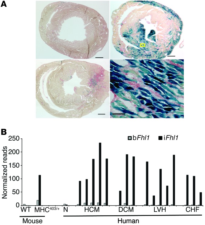
Figure 3. Fhl1 expression in mouse and human cardiomyopathic hearts.
(A) Histologic assessment of fibrosis and Fhl1 expression in male WT (Fhl1hemi/–) and HCM (Fhl1hemi/–) mouse hearts, assessed by lacZ expression in mice carrying the Fhl1lacZ allele (blue staining from β-gal with X-gal). Sections were also stained with Sirius red, which stains collagen in fibrotic areas red. The WT mouse expressed no iFhl1 and had no fibrosis. The young MHC403/+ mouse heart (bottom left) exhibited focal fibrosis (red) and iFhl1 expression (blue) prior to hypertrophy onset. An adult hypertrophic MHC403/+ mouse heart (top right) exhibited markedly increased iFhl1 expression in myocytes (bottom right, magnified view of boxed region) juxtaposed to interstitial fibrosis. Scale bars: 0.5 mm (left and top right); 0.05 mm (bottom right). (B) iFhl1 upregulation in mouse and human cardiomyopathies. Mouse 5′RNA-Seq libraries were constructed from RNA pooled from a minimum of 3 mouse hearts; human 5′RNA-Seq libraries were constructed from LV RNA extracted from individual subject hearts. N, normal; DCM, dilated cardiomyopathy; LVH, pressure overload LV hypertrophy; CHF, congestive heart failure.

