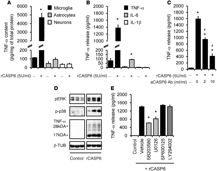
Figure 5. rCASP6 induces TNF-α release in microglial cultures via MAPK activation.
(A) TNF-α expression, revealed by ELISA analysis, in primary cultures of microglia, astrocytes, and DRG neurons after stimulation of rCASP6 (5 U/ml, 3 hours). *P < 0.05, n = 4 cultures. (B) Release of TNF-α, IL-1β, and IL-6 (ELISA analysis) in microglial culture medium after stimulation of rCASP6 (5 U/ml, 3 hours). *P < 0.05, compared with control, n = 4 cultures. (C) Dose-dependent inhibition of rCASP6-induced (5 U/ml, 3 hours) TNF-α release by CASP6-neutralizing antibody in microglial cultures. *P < 0.05, compared with control; #P < 0.05; compared with rCASP6, n = 3 cultures. (D) Expression of pERK, p-p38, and TNF-α, revealed by Western blot analysis, in microglial cultures after rCASP6 (5 U/ml, 3 hours) treatment. All the bands are from the same gel (Supplemental Figure 6E). Note a robust increase in the secreted form of TNF-α (17 kDa). (E) Effects of the MAPK inhibitor SB203580, U0126, and SP600125 (50 μM) and the PI3K inhibitor LY294002 (50 μM) on rCASP6-induced TNF-α release in microglial cultures. *P < 0.05, compared with vehicle (1% DMSO), n = 4 cultures.

