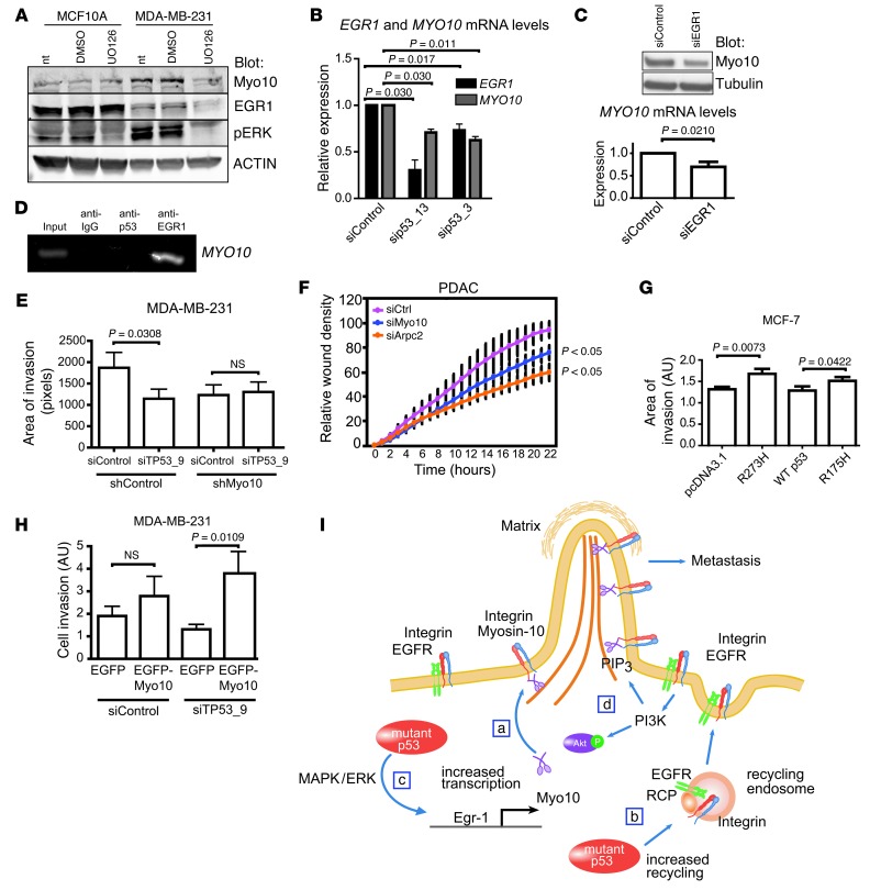
Figure 7. Myo10 is necessary for mutant p53–driven invasion.
(A) Western blot of MCF10A and MDA-MB-231 cells treated 16 hours with DMSO or MEK inhibitor (UO126, 10 μM). NT, non-treated. (B)TaqMan qRT-PCR of MYO10 and EGR1 mRNA levels in MDA-MB-231 cells silenced with 2 different p53 targeting oligos. n = 2 (sip53_13); n = 4 (sip53_3). (C) Western blot and TaqMan qRT-PCR showing Myo10 expression upon silencing of EGR1 for 48 hours. (D) ChIP analysis of mutant p53 or EGR1 binding to MYO10 promoter by PCR using Myo10 promoter–specific primers. IgG antibody was used as negative control. (E) Matrigel invasion of shMyo10 cells upon silencing of TP53 (areas of invasion, n = 10, ×20 objective). (F) Invasion of murine PDAC cells transfected with siMyo10 or siArpc2 (Arp2/3 component, positive control) into Matrigel-overlaid wounds. (G) MCF-7 cells were transfected as indicated, and invasion was analyzed as in E. (H) Invasion of p53-silenced MDA-MB-231 cells transfected as indicated (percentage of invaded/all GFP-positive cells; n = 9–14; ×20 objective. (I) Model of mutant p53–driven, integrin-dependent invasion. (a) Mutant p53 induces Myo10 that contributes to mutant p53–driven invasion by transporting integrin to filopodia tips. (b) Mutant p53 increases integrin and EGFR recycling to the plasma membrane (19) providing integrins to be transported to filopodia tips by Myo10. (c) Mutant p53 drives Myo10 by increasing EGR1 transcription directly and via MAPK/ERK signaling. (d) Increased integrin-EGFR recycling elevates PIP3 levels via PI3K/Akt pathway (19) and activates Myo10 (48). Mean ± SEM and Mann-Whitney test P values are shown.

