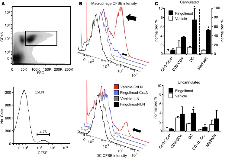
Figure 4. Leukocytes migrate from the CNS to CxLNs.
In icv-cannulated mice, after 7 days of CFSE infusion, CxLN CD45+ cells were gated and examined for CFSE labeling. (A) CFSE-labeled cells were discerned in all mice. The percentage of CFSE+ cells is indicated. (B) The same experiment repeated with fingolimod or vehicle administered i.p. (n = 6; 3 independent experiments), beginning 1 day prior to icv cannula implantation. For gating of CFSE+ CxLN and ILN monocytes, see Supplemental Figure 4. Top: Vehicle-treated mice (large arrow) had substantial populations of CFSE+ presumed macrophages (CD45+CD3–B220–CD11c–CD11b+), which were significantly reduced in CxLNs from fingolimod-treated mice (small arrow) and absent in ILNs of either vehicle- or drug- treated mice. Bottom: CFSE+ DCs (CD45+CD3–CD11c+) were only seen in CxLNs of vehicle-treated mice (arrow), and their accumulation was completely inhibited by systemic fingolimod treatment. (C) Mice that had CFSE infused by icv cannula had their brains removed, and mononuclear cells were extracted for FACS. Top: DC and macrophage/PMN accumulation in the brains of cannulated mice. Bottom: To determine which cell type(s) may have accumulated as a result of injury associated with icv cannulation, we also administered fingolimod (6 μg/d) or vehicle (n = 6; 3 independent experiments) i.p. to noncannulated mice, which demonstrated an increased frequency of DCs in the CNS as assessed by FACS (P = 0.018), with no differences in corresponding macrophage/PMN or T cell numbers. Data represent mean ± SEM. *P < 0.05.

