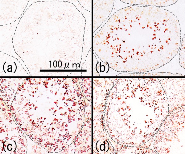
Fig. 5.

Cryostat sections of testes from normal mice (8 weeks of age). All the sections were reacted with 50-fold diluted serum samples obtained from the control group (a), TH group (b), TH+CFA+BP group (c) and CFA+BP group (d) followed by incubation with HRP-conjugated anti-mouse IgG antibody. The dashed line indicates the basal lamina of the seminiferous tubules.
