Fig. 5.

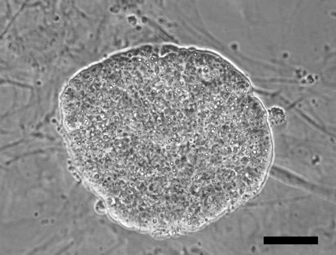
A bright field image of a primary colony generated by the trypsinization of a day 6 plated outgrowth showing an undifferentiated morphology. Scale bar is 50 µm.

A bright field image of a primary colony generated by the trypsinization of a day 6 plated outgrowth showing an undifferentiated morphology. Scale bar is 50 µm.