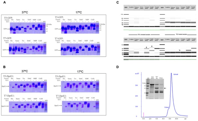
FIGURE 2.
Protein production screening in the vector suite. Panels (A,B) corresponds to the E-PAGE 96 acrylamide gels for the expression screening of GFP and DprE1, respectively. The incubation with TEV protease for fusion cleavage is indicated with a +sign over the corresponding lines. Cleaved target protein at the expected molecular weight (MW) is depicted. Additionally, induction temperatures are indicated over each panel. (C) Expression screening for MPK4 at 17°C using a Labchip GX II (Caliper, USA) microfluidic detection system. Arrows indicate the presence of a band with the expected molecular weight. Construct names are provided over each gel line. Solubility improvement with vector suite is indicated by arrows. (D) Analytical size exclusion chromatography (SEC) of the IMAC purified fraction of DsbC-MPK4. Peaks at 7.6 and 8.4 ml correspond to the exclusion volume and the 600 kDa decameric form of DsbC-MPK4, respectively. The 12% SDS-PAGE shows the fusion protein obtained by IMAC purification and DsbC-MPK4 digested by TEV protease. The expected molecular weight of MPK4 (41.7 kDa) is indicated by an arrow.

