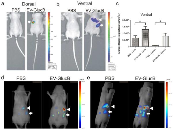
Figure 4. In vivo imaging of IV-administered EVs.
(a, b) Bioluminescence imaging of EV-GlucB in athymic nude mice. Animals were administered a bolus of either PBS (control) or EV-GlucB via the retro-orbital vein. CTZ was injected by the same route at 30 min post-initial administration to image EV-GlucB. (a) Representative image showing dorsal side of nude mouse with a prominent signal at regions corresponding to the spleen (arrow) in EV-GlucB-administered animals. (b) Imaging of ventral side showing a significant signal at regions corresponding to the spleen (arrow) and liver (arrowhead) in EV-GlucB-treated mice. No appreciable signal was detected on either side of PBS-injected mice. (c) Quantitation of EV-GlucB signal from bioluminescence imaging at ventral regions corresponding to the liver and spleen at 60 min post-EV administration. Sr, steradian. *, P < 0.05 by Student’s t-test. (d, e) FMT imaging of EV-GlucB in athymic nude mice. Alexa680-conjugated EV-GlucB or PBS (control) were administered via the tail vein and imaged with FMT at 30 min post-injection. (d) Single Z plane of FMT imaging showing elevated fluorescence signal predominantly at the spleen (arrow) and the liver (arrowhead) in Alexa680-EV-GlucB, but not in control-treated mice. (e) 3D representation of FMT imaging illustrating Alexa680-EV-GlucB localizing mainly to the spleen (arrow) and the liver (arrowhead). A low level of background signal was also detected in the control.

