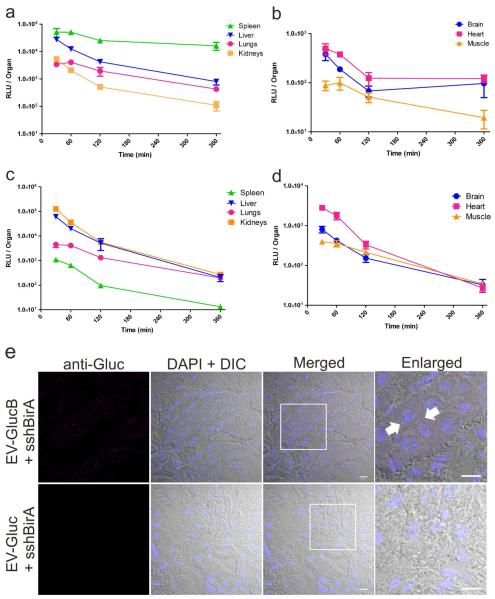
Figure 5. Biodistribution and retention of IV-administered EV-GlucB.
(a, b) Biodistribution of IV-injected EV-GlucB via tail vein over time. Gluc activity was measured from organs collected from mice at different time points following EV-GlucB injection without transcardial perfusion with PBS. (c, d) Tissue retention of IV-injected EV-GlucB via tail vein over time in perfused tissues. Organs were collected from EV-injected animals at the same time points as above following transcardial perfusion with PBS. (e) Subcellular visualization of EV-GlucB in perfused kidneys by confocal microscopy. Kidney samples were collected from mice 30 min after IV-administration of EV-GlucB or EV-Gluc and transcardial perfusion with PBS, then cryosectioned and immunostained with anti-Gluc (rabbit) and Alexa Fluor® 647 goat anti-rabbit antibodies. EV-GlucB (arrow) was detected in a punctuate pattern in the perinuclear region of renal cells. EV-Gluc was used as a negative control for nonspecific binding or packaging of secreted Gluc protein in EVs and unspecific binding of the anti-Gluc antibodies to EVs. Nuclei were visualized by 4,6-diamidino-2-phenylindole (DAPI). DIC, differential interference contrast. Bar, 10 μm.

