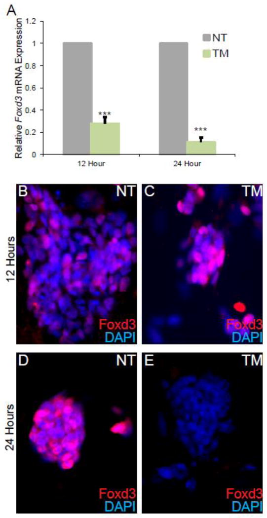
Figure 1. Foxd3 protein cannot be detected after 24 hours in culture with Tamoxifen (TM).
A. qRT-PCR analysis of Foxd3 mRNA levels after 12 and 24 hours of culture with TM. Relative Foxd3 expression is decreased in TM-treated ESCs (green) at both time points compared to untreated controls (grey). Error bars indicate SEM. *** p<0.001. N=3 experiments. The expression of Foxd3 in NT cells is set to 1. B–E. Immunocytochemistry analysis of Foxd3 protein expression (red) after 12 (B–C) and 24 (D–E) hours in culture in NT (B,D) and TM-treated (C,E) ESCs. Nuclei are indicated by DAPI (blue).

