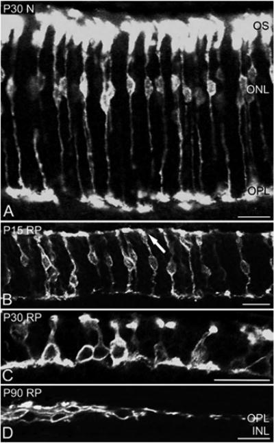
Figure 1.

Confocal micrographs taken from vertical sections of retinas processed for M-opsin immunoreactivity. The micrographs are for P30 N (A), P15RP (B), P30RP (C), and P90 RP (D). In P30 N retinas, entire M-opsin-immunoreactive cones are labeled. In P15 RP retinas, the OS are distorted in orientation (arrow). In P30 RP retinas, M-opsin immunoreactive cones are shortened in length and show disorganized axon terminals (C). In P90 RP retinas, M-opsin-immunoreactive cones are positioned “flat” against the outer part of the INL. ONL, outer nuclear layer; OPL, outer plexiform layer; INL, Inner nuclear layer; OS, outer segment; N, normal; RP, retinitis pigmentosa. Scale bars = 20 μm.
