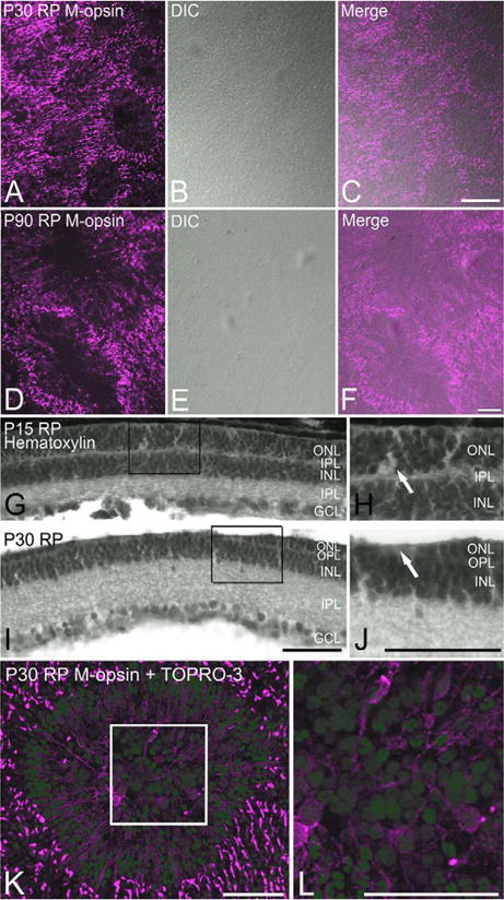
Figure 3.

Confocal micrograph taken from P30 (A–C) and P90 (D–F) whole mount RP retinas processed for M-opsin immunoreactivities showing rings in their distribution (A,D). Light micrograph taken at the same retinal location under DIC mode shows no retinal folds (B,E). Double exposure (C,F) confirms no retinal folds are associated with rings. Light micrographs taken from RP vertical retinas processed with hematoxylin staining (G–J). At P15, the thickness of the ONL is uniform (G). H: Higher-power micrograph of G is shown. At P30, the ONL show “grooves.” J: Higher-power micrograph of groove is shown. No nuclei are visible at the trough of the groove (arrow). Confocal micrograph taken from P30 whole mount RP retina processed with M-opsin antibody (red) and TOPRO-3 (blue). Nuclei at the center of the ring are not in the same focal plane as M-opsin-immunoreactive cones (K). L: Higher-power micrograph of K is shown. DIC, differential interference contrast; ONL, outer nuclear layer; INL, inner nuclear layer; IPL, inner plexiform layer; GCL, ganglion cell layer. Scale bars = 100 μm in A–J; 50 μm in K,L. [Color figure can be viewed in the online issue, which is available at wileyonlinelibrary.com.]
