Figure 4.

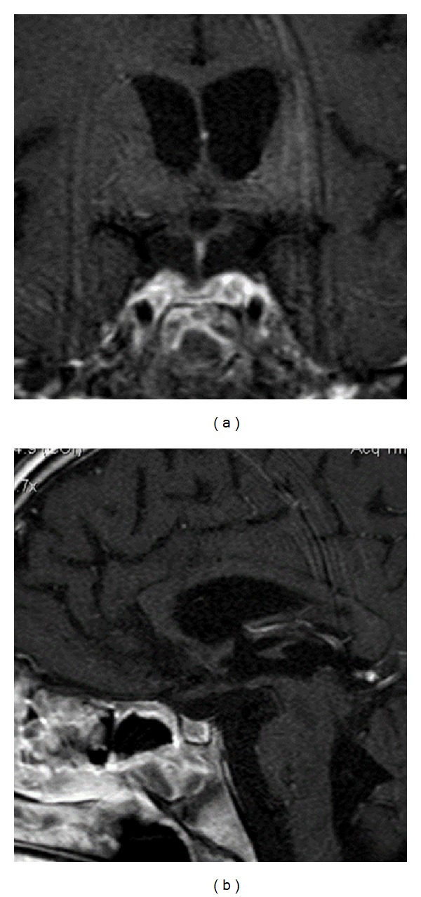
Axial T1-weighted (a) and sagittal T1-weighted (b) MRI after gadolinium enhancement one month after chemotherapy, MRI showed resolution of pituitary-hypothalamic mass, with a marked reduction in stalk-thickness.

Axial T1-weighted (a) and sagittal T1-weighted (b) MRI after gadolinium enhancement one month after chemotherapy, MRI showed resolution of pituitary-hypothalamic mass, with a marked reduction in stalk-thickness.