
Figure 3.
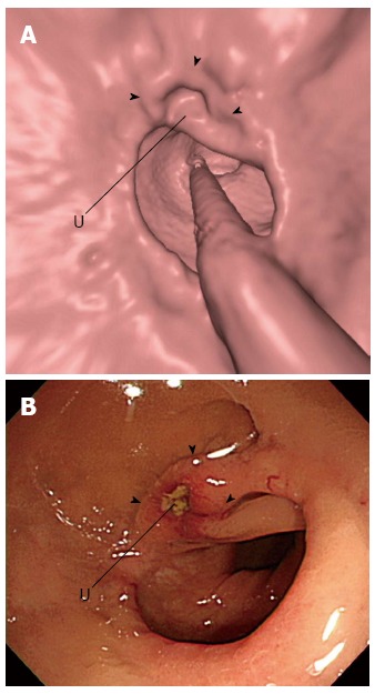
A 66-year-old man with a 16-mm ulcerating anastomotic recurrence. Three-dimensional endoluminal computed tomographic colonographic (A; U = Ulcer) and colonoscopic (B; U = Ulcer) images obtained 10 mo after cancer resection surgery show an ill-defined elevated lesion with central ulceration (arrowheads) at the anastomosis site. Subsequent surgical resection and pathologic analysis confirmed recurrent adenocarcinoma (reprint with permission[68]).