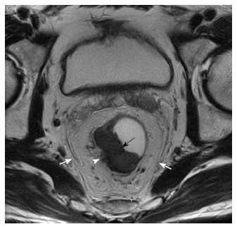
Figure 2.

T3 tumor, circumferential resection margin not threatened. T2W axial magnetic resonance imaging image shows a mildly hyperintense proliferative tumor along the right lateral and posterior wall (black arrow). Arrow head shows the tumor reaching upto the muscularis with spiculation in the adjacent perirectal fat. The white arrows show the mesorectal fascia which is not involved/threatened.
