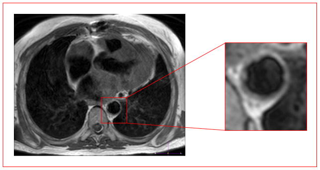
Figure 1.

Pericardial fat can be expanded in otherwise lean but metabolically unhealthy subjects. MRI images of a metabolically unhealthy, lean (MUL) subject, showing ectopic fat deposition consistent with previous reports of ectopic adipose tissue accumulation [27,28] in such subjects. An axial T1-weighted black-blood image obtained through the thorax shows pericardial fat (bright regions surrounding the heart) and aortic atherosclerosis (shown in the red box enlargement) (irregular thickening of the vessel wall) in a non-obese subject with metabolic syndrome. There is also prominent fat surrounding the coronary above the aorta.
