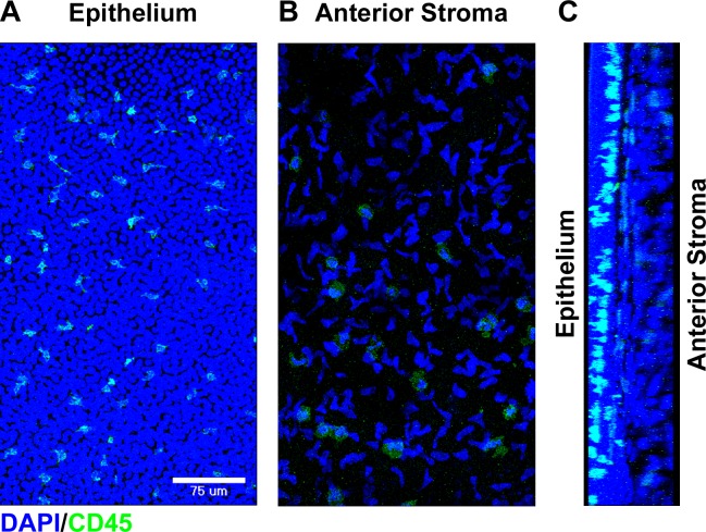
Figure 1.
Bone marrow–derived cells within the normal human cornea. Fresh donor human corneal tissue was fixed, and whole mounts were stained with the indicated markers prior to confocal imaging. Compressed images of z-stacks in the xy plane from the peripheral corneal epithelium (A) and anterior stroma (B) showing staining for DAPI (blue) and CD45 (green). (C) Cross-sectional reconstruction in the yz plane (epithelium on the left and anterior stroma on the right) of the peripheral cornea showing staining for DAPI (blue) and CD45 (green).

