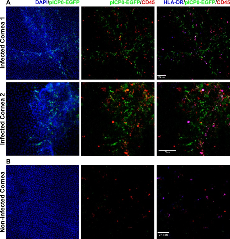
Figure 4.
HSV-1 infection of the corneal epithelium induces migration of tissue-resident APCs. (A) The epithelium of human corneal tissue was scarified and pICP0-EGFP HSV-1 RE applied. The tissue was incubated at 37°C for 1 hour and then washed extensively. The tissue was placed in fresh media and further cultured for various times (16 hours depicted in the figure). The tissue was then fixed and stained with the indicated markers prior to confocal imaging. Compressed images of z-stacks in the xy plane from the peripheral epithelium and anterior stroma of two separate corneas are shown. Epithelial scarification and infection lead to disruption of the normal anterior corneal architecture as assessed by DAPI staining. APCs were found throughout the remaining epithelium and anterior stroma. Data are representative of three separate corneas. (B) Tissue that was not scarified or infected but otherwise treated as above served as control. Compressed image of a z-stack in the xy plane from the peripheral epithelium is shown.

