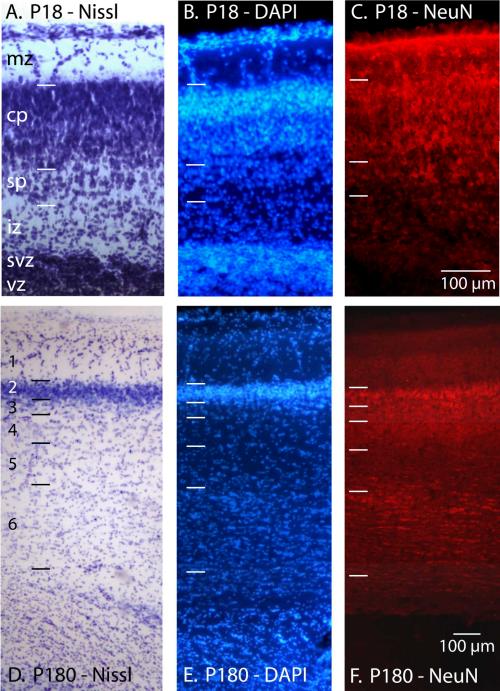
Figure 6.
The normal, anisotropic distribution of cells in cortical tissue stained for Nissl (A, D), DAPI (B, E), and NeuN (C, F) in P18 (A-C) and P180 (D-F) opossums. Both the P18 and P180 cortical sections are from a similar location in somatosensory cortex. At P18 the laminar organization of the developing neocortex is distinct and neurons within the cortical plate are clearly labeled with NeuN. While the ventricular zone is cell dense it contains no NeuN labeled cells. In the adult the characteristic layers of the neocortex are visible in both Nissl and DAPI stained tissue. Tissue stained for NeuN indicates a lack of neurons in layer 1, dense labeling of neurons in layers 2-3, and moderate labeling of neurons in layer 4 and 6. mz = marginal zone; cp = cortical plate; sp=subplate; iz = intermediate zone; svz = subventricular zone; vz = ventricular zone. Laminar division of early postnatal animals have been described by (Cheung et al., 2010; Saunders et al., 1989). Color and contrast have been adjusted using Adobe Photoshop. The scale for all images = 100 μm.

