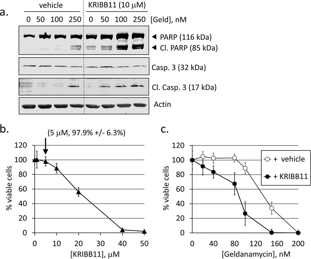
Fig. 3.
Biochemical inhibition of HSF1 activity or inhibition autophagy both sensitize cells to geldanamycin-induced apoptotic cell death. a. RKO were pre-treated with vehicle (0.1% DMSO) or 10 nM KRIBB11 for 1 h followed by geldanamycin (50–250 nM) for 24 h. Total protein extracts were analyzed for PARP and caspase-3 cleavage. b. RKO were treated with KRIBB11 (1–50 nM) for 48 h. Data points represent mean values of Calcein-AM fluorescence normalized to vehicle-treated (0.1% DMSO) control. Error bars are standard deviations for n = 8. Label indicates % viability vs. control for 5 nM KRIBB11) c. Concentration-response curves for cell viability in RKO treated with geldanamycin (20–200 nM) + 0.1% DMSO (vehicle control, open circles) or geldanamycin (20–200 nM) + 5 nM KRIBB11. Error bars are standard deviations for n = 8.

