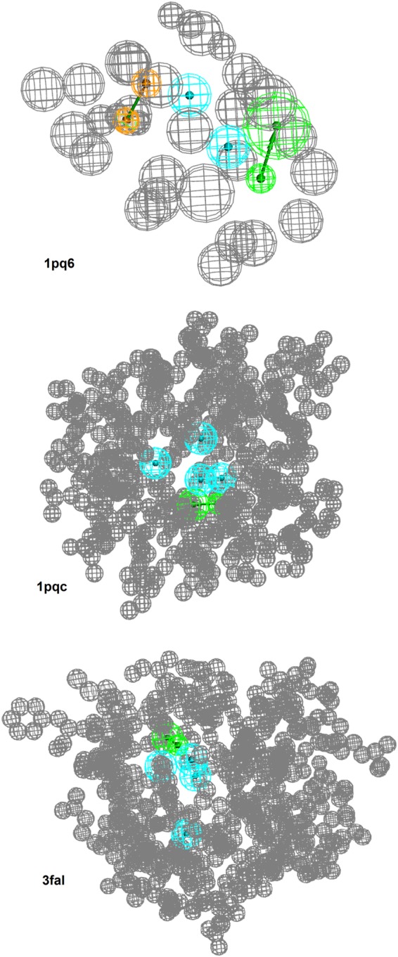
Figure 1.

Pharmacophore models that showed a significant enrichment of highly LXRβ-selective compounds. The models are named after the X-ray crystal structure protein data bank22 code from which they were originally derived. Blue spheres illustrate hydrophobic features. Green arrows represent hydrogen bond acceptors. Brown spheres represent aromatic interactions with indicated direction. The gray spheres signify so-called exclusion volumes that represent the space occupied by the protein. Model 1pc6 consists of two hydrophobic features, one hydrogen bond acceptor, an aromatic interaction, and 31 exclusion volumes. Model 1pqc consists of three hydrophobic features, one hydrogen bond acceptor, and 458 exclusion volumes. Model 3fal consists of three hydrophobic features, one hydrogen bond acceptor, and 514 exclusion volumes.
