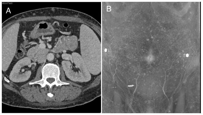
Figure 1. Radiographic studies of the abdomen and pelvis demonstrate numerous subcutaneous nodules.

Axial computed tomography scans show multiple nodules in the subcutaneous fat without any enhancement (A). The 3D volume rendered images show the extent of lesion involvement over the abdominal wall (B). These nodules also demonstrated mild FDG uptake (not shown).
