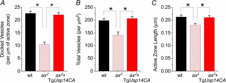
Figure 7.
Transmission electron micrographs of lateral perforant path synapses from wt mice, axJ mice and axJ + TgUsp14CA mice were analysed. There is a decrease in docked vesicles (A), total vesicles (B), and active zone length (C) at lateral perforant path synapses from axJ mice (n = 69) compared to synapses from wt controls (n = 65), P < 0.01. All three measures are restored at synapses from axJ + TgUsp14CA mice (n = 65). *P < 0.05.

