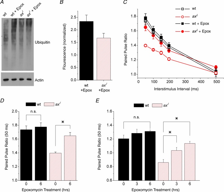
Figure 8.
A, immunoblot for ubiquitinated proteins isolated from hippocampal slices after 6 h of proteasome inhibition with epoxomycin (10 μm). B, quantification of immunoblots as in A, n = 5 wt, n = 5 axJ. Results are normalized to fluorescence from slices with no epoxomycin. C, paired pulse facilitation measured at Schaffer collateral synapses in slices from wt mice (n = 4) and axJ mice (n = 5) after 6 h of exposure to epoxomycin or vehicle. axJ slices showed a robust increase in PPF while wt slices showed no change. D, paired pulse facilitation at Schaffer collateral synapses at the 50 ms interval for 0 and 6 h treatment with epoxomycin (10 μm), n = 4 wt, n = 5 axJ. *P < 0.05. E, paired pulse facilitation at lateral perforant path synapses at the 50 ms interval for 0, 3 h and 6 h treatment with epoxomycin (10 μm), n = 4 wt, n = 4 axJ. *P < 0.05.

