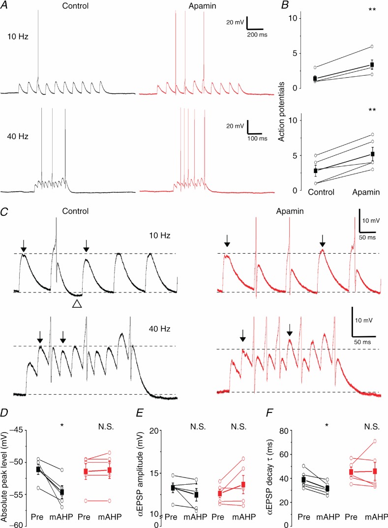
Figure 9.
A, representative examples showing the effect of SK channel blockade on subthreshold summation recorded at 36°C. αEPSPs were injected at 10 and 40 Hz into the somatic compartment while keeping the neuron depolarized near AP threshold. B, SK channel blockade significantly increased the number of spikes during a 10 αEPSP train both at 10 and 40 Hz (n = 5, P < 0.01 (**)). C, examples shown in A at higher magnification clearly show the reduced summation of αEPSPs during the mAHP and the dramatic increase after apamin application. Top arrows show αEPSPs before and during the mAHP. Note the undershoot due to the mAHP (open triangle). D, E and F, summary plots comparing the absolute peak level, amplitude and decay time constant (τ) of the αEPSP before the first AP (‘Pre’) and the first αEPSP after the first AP and associated mAHP (‘mAHP’) in control and apamin conditions. The mAHP significantly reduced the absolute αEPSP peak (D) and decay time constant (τ) (F) (n = 5, P < 0.05 (*)) under control conditions but apamin abolished this difference.

