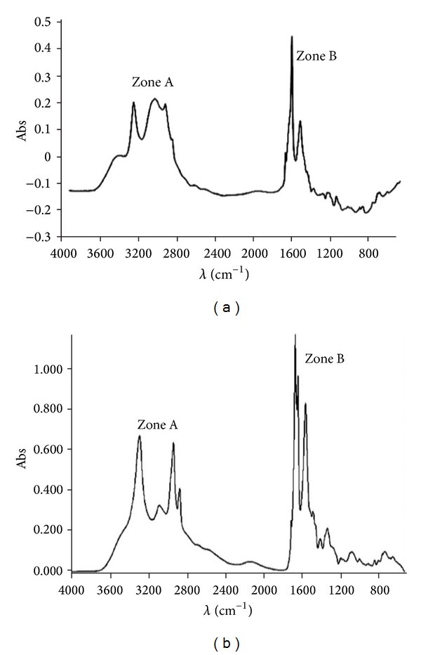
Figure 2.

Infrared spectra of poly-L-lysine (a) and lauryl-PLL (b). Fatty acid-PLL compounds were analyzed by ATR-FTIR spectroscopy (with a Bruker apparatus). Zone A: in free PLL (a), 3,200 to 2,850 cm−1, CH2 groups of side chains of amino acid lysine. In lauryl-PLL (b), CH3 groups at the end of the lauric acid chain are not very visible. Note that there is a weak shoulder the CH2 peak at 2,925 cm−1. Zone B: in free PLL (a), 1,700 to 1,450 cm−1, deformation bands of the amino group (2), NH, and of carbonyl groups, CO. In lauryl-PLL (b), these two peaks increase in intensity because of additional CO and NH by the linkage of fatty acid.
