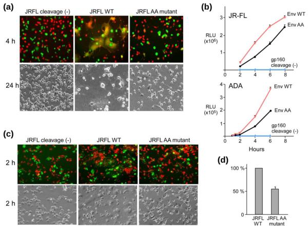
Fig. 4.
Env-mediated fusion impaired by AA mutations. (a) Qualitative microscopy analysis of cell-cell fusion. Content mixing of Env-expressing 293T effector cells with 3T3.CD4.CCR5 target cells. The top row shows the overlay of fluorescence images after co-incubation of 293T cells (Green, Calcein) with 3T3.CD4.CCR5 cells (Red, CMTMR) at 37°C for 2 h. The bottom row shows representative bright-field images collected 24 h after co-incubation at 37°C. (b) Fusion kinetics of JR-FL (top) or ADA (bottom) Env-expressing 293T cells with TZM-bl cells containing a Tat-driven luciferase reporter. (c) Hemifusion identified by lipid mixing between 293T cells (green) and DiI-labeled 3T3.CD4.CCR5 cells (red). The top row shows the overlay of fluorescence images collected 2 h after co-incubation at 37°C. The bottom row shows the corresponding bright field images. (d) Quantitative lipid mixing efficiency of JRFL-AA relative to WT. Flow-cytometric analysis of lipid-dye transfer between DiO labeled 293T cells and DiD labeled TZM-bl cells were conducted. The percentage lipid mixing activities were determined following the subtraction of background dye redistribution between empty vector-transfected effector and target cells, normalized to that of WT (100%) in three independent experiments.

