
Figure 2.
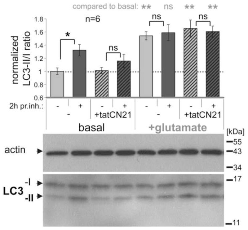
Autophagic flux in hippocampal cultures is inhibited by tatCN21 and by glutamate, as revealed by the LC3-II/I immuno-detection ratio with or without a 2 h treatment with protease inhibitors (pr. inh.). Shown are example Western-blots (lower panel) and their quantification (upper panel; n=6) normalized to the basal LC3-II/I ratio (which was ~0.5). Under basal conditions, protease inhibitor treatment significantly increased the LC3-II/I ratio, due to block of autophagic degradation of LC3-II. This increase was reduced by tatCN21 (5 μM) and completely blocked by excitotoxic glutamate (100 μM for 5 min). In comparison of different conditions without protease inhibitors, glutamate treatment increased the LC3-II/I ratio (indicating a late stage block of autophagic flux) while tatCN21 treatment did not (indicating instead an early stage inhibition of autophagy). **: p<0.01; *: p<0.05 in ANOVA with Newman-Keuls post-hoc analysis; ns: p>0.05 even in one-tailed t-test. Bar graphs indicate mean±SEM.