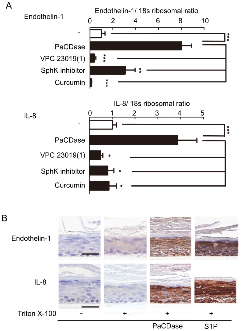
Figure 5. PaCDase-produced S1P induces endothelin-1 and IL-8 production by keratinocytes.
(A) Involvement of SphK and S1P receptor in PaCDase-enhanced endothelin-1 and IL-8 gene expression. Nitrocellulose filters without (-) or with 1 mU/ml PaCDase in the absence or presence of 1 µM VPC 23019, 10 µM SphK inhibitor, or 40 µM curcumin in Tris-buffered saline containing 0.1% Triton X-100 were placed on the stratum corneum, and endothelin-1 and IL-8 mRNAs were assayed by quantitative real-time RT-PCR. Each bar represents the mean±SD of 3 independent experiments. **P<0.01; ***P<0.001. (B) Immunohistochemical analysis. Nitrocellulose filters with Tris-buffered saline alone (-/-), or without (+/−) or with 1 mU/ml PaCDase (+/PaCD) or 5 µM S1P (+/S1P) in Tris-buffered saline containing 0.1% Triton X-100 were placed on the stratum corneum. The cells were incubated for 24 h, embedded in paraffin, sectioned, and incubated with rabbit anti-endothelin-1 IgG (endothelin-s) or mouse anti-human IL-8 IgG. The data shown represent 3 independent experiments. Bar: 25 µm.

