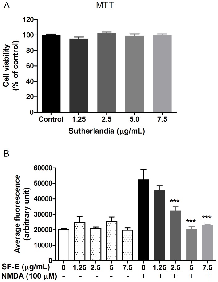
Figure 1. SF-E inhibits NMDA-induced ROS production without altering cell viability of primary cortical neurons.
(A) Exposure of SF-E (0–7.5 µg/mL) to primary cortical neurons for 24 h did not alter neuronal viability as assayed by MTT. Data are expressed as the mean ± SEM from 3 individual experiments and analyzed by one-way ANOVA (p = 0.1774). (B) Bar graph of average fluorescence, depicting inhibition of NMDA-induced ROS production by SF-E. ROS production was determined in primary neurons after treating cells with SF-E (0–7.5 µg/mL) for 30 min prior to stimulation with NMDA (100 µM) for 30 min. For ROS production, neurons were loaded with dihydroethidium (DHE, 10 µM) 30 min prior to image acquisition. Data are expressed as the mean ± SEM from 3 individual experiments and analyzed by two-way ANOVA with Bonferroni post-tests. ***indicates significant decrease in ROS production by SF-E as compared to NMDA (p<0.001).

