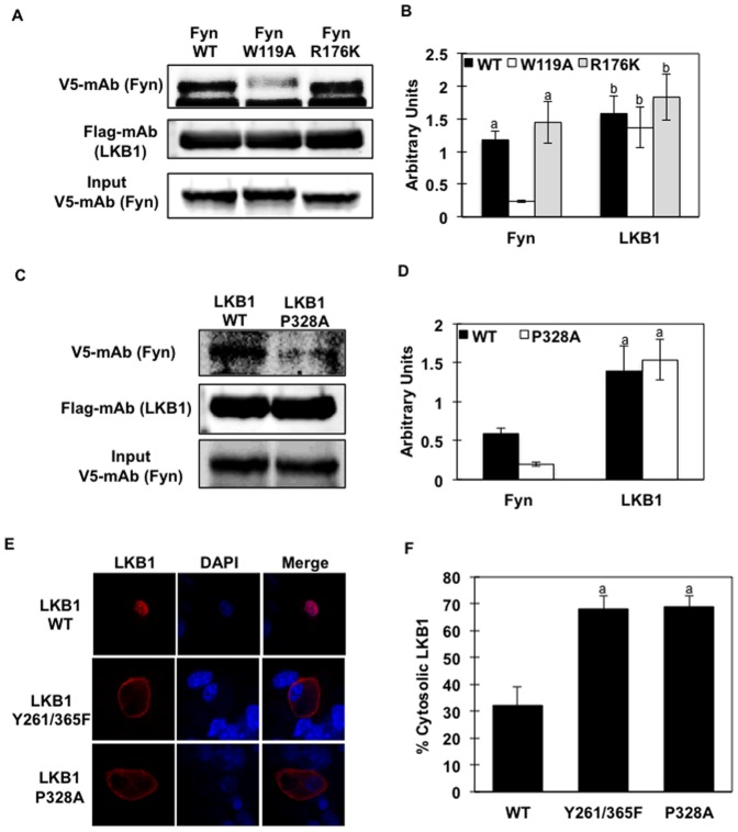
Figure 2. Fyn SH3 domain binds to the proline-rich domain of LKB1 and induces LKB1 cytosol localization in vivo (A) Lysates from HeLa cells expressing Flag-LKB1-WT were immobilized onto Flag-conjugated beads.
Protein extracts (same amount) from HeLa cells expressing V5-Fyn-WT or V5-Fyn-W119A or V5-Fyn-R176K constructs were incubated with the Flag-LKB1-WT. Proteins were separated by electrophoresis and immunoblotting was performed using specific V5 antibody (detecting Fyn) and Flag antibody (detecting LKB1). (B) Signal quantification from 3 independent experiments. Identical letters indicate values that are not statistically different from each other (P>0.05). (C) Lysates from HeLa cells expressing either Flag-LKB1-WT or Flag-LKB1-P328A were immobilized onto Flag-conjugated beads. Protein extracts (same amount) from HeLa cells expressing V5-Fyn-WT construct were incubated with either Flag-LKB1-WT or Flag-LKB1-P328A. Proteins were separated by electrophoresis and immunoblotting was performed using specific V5 antibody (detecting Fyn) and Flag antibody (detecting LKB1). (D) Signal quantification from 3 independent experiments. Identical letters indicate values that are not statistically different from each other (P>0.05). Images in (A and C) are representative of 3 independent experiments. (E) Fully differentiated 3T3L1 adipocytes were co-transfected with pcDNA3-Flag-LKB1 or pcDNA-LKB1-P328A. LKB1 subcellular localization was assessed by immunofluorescence using the rabbit Flag polyclonal antibody followed by Alexa Fluor 488 Anti-Rabbit IgG. Images are representative of 3 independent experiments. (E) Percentage of 3T3L1 cells with pcDNA-Flag-LKB1 signal detected in the cytoplasm. Data are representative of 3 independent experiments.

