
Fig. 1. Pulmonary vaccination increases DC uptake of nanocapsules, enhances T cell priming, and promotes imprinting of mucosal homing receptors.
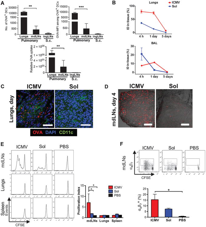
C57BL/6 mice were immunized subcutaneously or intratracheally with OVA in either soluble or ICMV formulations with MPLA and polyI:C adjuvants. (A) Numbers of DCs containing OVA and the MFI of OVA in individual cells were measured in the lungs and mdLNs on day 2 after intratracheal administration. Numbers were compared to those in ingLNs after subcutaneous (s.c.) administration. Relative OVA uptake was calculated by multiplying OVA+ DC counts by mean OVA-MFI. (B) The amount of antigen remaining in the lungs and BAL was measured over time by fluorescence spectroscopy. ID, injected dose. (C and D) Representative cryosections after intratracheal immunization with fluorescent OVA (red) from lungs on day 1 (C; scale bars, 50 μm) and mdLNs on day 4 (D; scale bars, 200 μm). Lung sections were costained with anti-CD11c and 4′,6-diamidino-2-phenylindole (DAPI). (E and F) Tissues harvested 3 days after intratracheal immunization were homogenized, and recovered cells were cocultured with CFSE-labeled OT-I CD8+ T cells. (E) The proliferation index was calculated as the total number of cell divisions divided by the number of cells that went into division. (F) Expression of α4β7 on OT-I CD8+ T cells determined by flow cytometry after 3 days of coculture. Data are means ± SEM of two to three independent experiments conducted with n = 3 to 4 animals per group. *P < 0.05, **P < 0.01, ***P < 0.001, by one-way analysis of variance (ANOVA) (A and F) or two-way ANOVA (E).