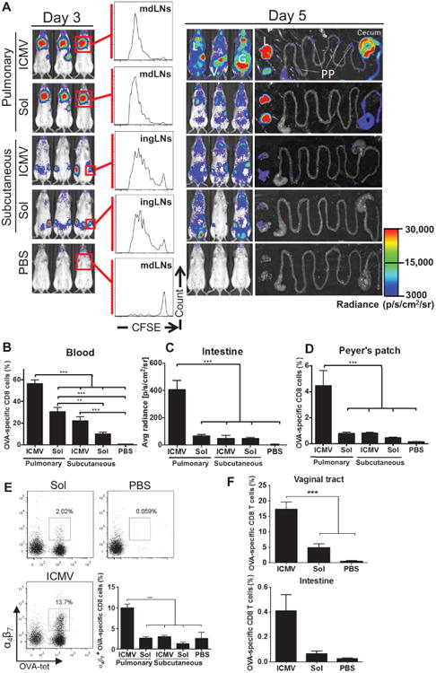
Fig. 3. Pulmonary ICMV immunization induces dissemination of CD8 T cells from mdLNs to distant mucosal tissues.

(A to E) OT-I–luc CD8+ T cells were adoptively transferred into C57BL/6 mice (n = 5) 1 day before ICMV or soluble OVA vaccine immunization via intratracheal or subcutaneous routes. (A) Trafficking and proliferation of OT-I–luc T cells were monitored by flow cytometry and bioluminescence imaging on days 3 and 5 after immunization. Flow cytometry histograms show representative CFSE dilutions in transferred T cells on day 3 after immunization at dLNs (mdLNs for intratracheal vaccines and ingLNs for subcutaneous vaccinations). Lungs (L) and gastrointestinal tracts (G) were dissected on day 5 and imaged to identify T cell localization. V, vaginal tract; PP, Peyer's patches. (B to E) Flow cytometry and imaging analyses of OT-I T cells on day 5. Shown are frequencies of OVA-specific CD8+ T cells in blood (B) and Peyer's patches (D), quantification of bioluminescence signal from small intestines (C), and integrin α4β7+ cells (E) in blood. (F) C57BL/6 mice (no adoptive transfers, n = 3 per group) were immunized with ICMV or soluble OVA vaccines on days 0 and 28. The frequency of OVA-specific CD8+ T cells in the vaginal tract and in the small intestine on day 7 after boost was assessed by flow cytometry. Data are means ± SEM of two independent experiments. **P < 0.01, ***P < 0.001, by one-way ANOVA.
