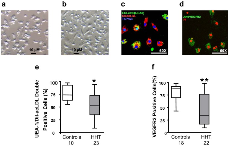
Figure 2. Reduced development of endothelial cellular phenotypes in CACs from HHT patients.
CACs derived from patients with HHT appeared smaller in size and more rounded (a) when compared to the spindle-shaped, elongated morphology of CACs derived from control subjects (b). Dil-Ac-LDL/UEA-1 double positive cells (a representative image shown in c. Orange: double positive; green: UEA-1 positive; red: Dil-Ac-LDL positive; blue is nuclear stain) and VEGFR2 positive cells (a representative image shown in d. Green: VEGFR2 positive; red is nuclear stain) in CACs were analyzed by immunofluorescence microscopy. The results showed that percentage of Dil-Ac-LDL/UEA-1 double positive cells and percentage of VEGFR2 positive cells, both were significantly lower in CACs isolated from HHT patients (e and f). *p<0.05, compared with healthy controls. **p<0.01, compared with healthy controls.

