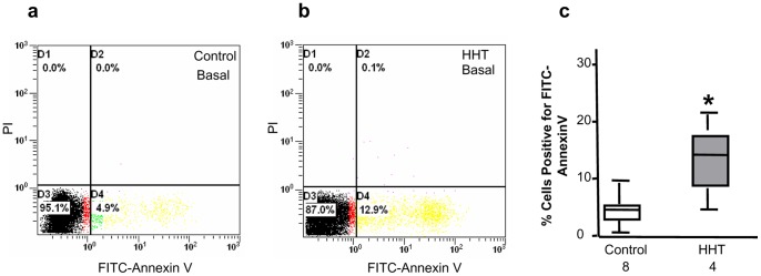
Figure 3. Elevated basal cell apoptosis levels in CACs from HHT patients.

Basal cell apoptosis in CACs after a 7-day culture was analyzed by Annexin V staining and flow cytometry. A representative dot plot of flow cytometric analysis for healthy and HHT CAC apoptosis was shown in panels a and b, respectively. Statistic analysis demonstrated that the levels of basal cell apoptosis in CACs from HHT patients were significantly higher than those in CACs of healthy individuals (c). *p<0.05, compared with healthy controls.
