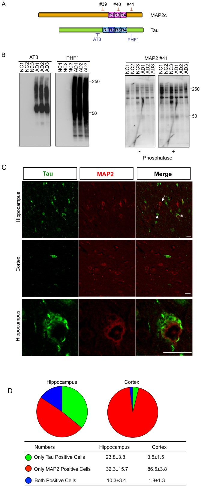
Figure 6. MAP2 is not involved in the growth process of NFTs in the AD brain.
(A) Diagram of three site-specific anti-MAP2 antibodies and anti-Tau antibodies. (B) Human temporal cortex tissues from three AD patients and three normal controls were homogenized sequentially in detergent-containing buffers. Sarkosyl-insoluble, SDS-soluble fractions were prepared and subjected to SDS-PAGE followed by western blotting using anti-Tau antibodies (AT8 and PHF1) and three newly raised site-specific anti-MAP2 antibodies (MAP2-#39 and #40 are not shown). Total protein was used as the loading control. The staining intensity of Tau was increased markedly in the Sarkosyl-insoluble, SDS-soluble fractions from AD brains compared with normal brains. By contrast, the MAP2 antibodies failed to detect any increased patterns in AD brains compared with normal brains. Phosphatase treatment was performed to avoid effects from MAP2 phosphorylation. NC, normal brains; AD, Alzheimer's disease brains. Information about the cases is provided in Table S1. (C) Double immunofluorescence staining of the homologous carboxyl-terminal sequences of Tau and MAP2 in the AD brain. AD brain paraffin-embedded sections were double-labeled by anti-Tau antibody (PHF1) and anti-MAP2 antibody (MAP2-#41). Tau but not MAP2 localized in NFTs as well as in NTs. Representative Tau-positive-only neurons (arrowhead), MAP2-positive-only neurons (arrow) and Tau/MAP2-double-positive neurons (star) are indicated. Scale bars = 25 µm. (D) Average number of the three neuron types was counted per 640 µm2. The data are presented as the mean±SD.

