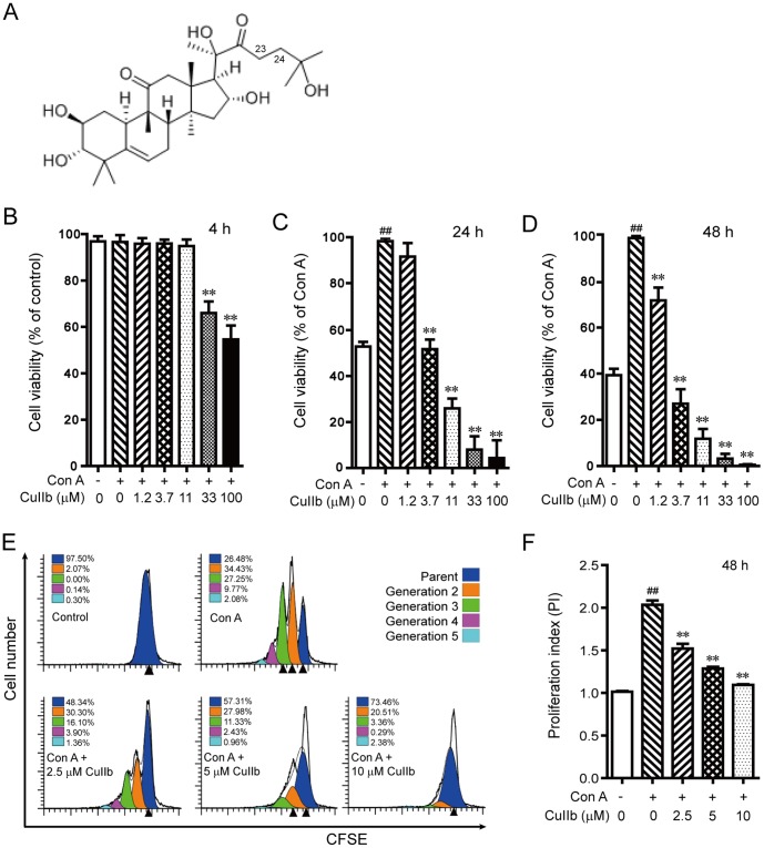
Figure 1. The chemical structure of cucurbitacin IIb (CuIIb) (A) and its effect on the proliferation of Con A-stimulated lymphocytes.
Mouse lymphocytes were incubated with different concentrations of CuIIb for 4 h, 24 h or 48 h, respectively and analyzed with WST-1 assay (B, C and D). Cell division was also measured by CFSE staining assay (E) and the proliferation indexes of CFSE-labeled cells are presented in (F). Data are analyzed with ModFit software. One representative data of three independent experiments with similar results are shown as mean ± SD (n = 3). ##P<0.01 versus control group; **P<0.01 versus Con A group.

