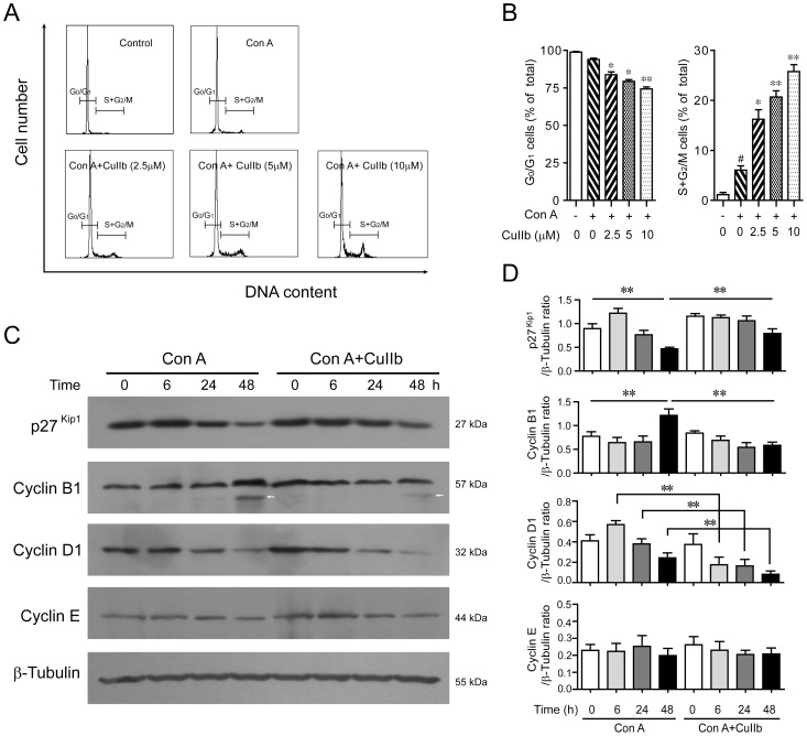
Figure 2. CuIIb induced cell cycle arrest by modulating the expression of cell cycle-related proteins in activated lymphocytes.
(A and B) Flow cytometry analysis showing cell cycle distribution of Con A-stimulated lymphocytes upon CuIIb treatment for 48 h. #P<0.05 versus control group; *P<0.05 and **P<0.01 versus Con A group. (C and D) Cells were pretreated with CuIIb (10 µM) for 1 h, then exposed to Con A (5 µg/ml) for 0, 6, 24, and 48 h, respectively. The expression of p27Kip1 and cyclin proteins at various time points was determined by Western blotting. β-Tubulin was used as a loading control. Representative blots of three independent experiments are shown in (C) and the relative densitometric ratios of each protein to β-tubulin are shown in (D). Values are shown as mean ± SD of three experiments. Arrow indicates a nonspecific band. **P<0.01.

