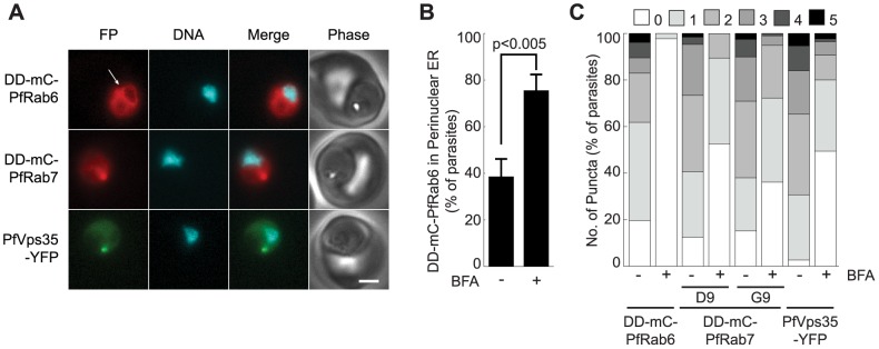
Figure 3. PfRab6 but not PfRab7 or retromer rapidly redistributes to the ER upon brefeldin A treatment.

(A) Distributions of DD-mCherry-PfRab6, DD-mCherry-PfRab7 and PfVps35-YFP in live parasites after one hour in the presence of 5 µg/mL brefeldin A. Redistribution of DD-mCherry-PfRab6 to the perinuclear ER is indicated with an arrow (top panel). FP, fluorescent protein. mCherry fluorescence is pseudocolored red, YFP fluorescence is pseudocolored green and Hoechst 33342 fluorescence is pseudocolored cyan. Scale bar, 2 µm. (B) Effect of brefeldin A (5 µg/mL, 1 hour) on the percentage of parasites exhibiting ER-associated DD-mCherry-PfRab6 fluorescence. Results are an average of three experiments, n = 75 to 85 parasites per condition. The p-value was determined using a two-tailed Student's t-test. (C) Effect of Brefeldin A (5 µg/mL, 1 hour) on the number of puncta labeled with DD-mCherry-PfRab6, DD-mCherry-PfRab7 (clones D9 and G9) or PfVps35-YFP. Results are averages of three experiments, n = 50 to 70 parasites per condition.
