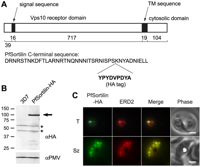
Figure 4. The putative protein sorting receptor PfSortilin localizes to the P. falciparum Golgi apparatus.
(A) Schematic diagram of the domain organization of PfSortilin with the number of amino acids in each domain indicated below. At bottom is the C-terminal sequence of PfSortilin with the site of incorporation of the HA tag indicated. TM, transmembrane. (B) Anti-HA immunoblot of the membrane fraction of parasites expressing PfSortilin-HA (clone C9) and of the parental 3D7 parasite line. PfSortilin-HA is indicated with an arrow. Two cross-reacting species are present in both lanes (asterisks). The membrane was reprobed with anti-plasmepsin V (PMV) antibodies for a loading control. The sizes of protein markers in kDa are indicated at left. (C) Co-localization of PfSortilin-HA and ERD2 in aldehyde-fixed clone C9 parasites. T, trophozoite; Sz, schizont. Scale bar, 2 µm.

