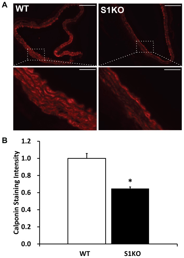
Figure 6. Reduced expression of calponin by aged syndecan-1 knockout (S1KO) mouse aortae relative to old wild type (WT) mice aorta.

(A) Immunohistochemical staining for calponin in the descending aorta harvested from the mice. (B) Morphometric quantification of calponin staining in the arteries (n = 5). All images were taken at the same length of exposure and calponin staining relative intensities were quantified. The intensities for S1KO mouse descending aorta were normalized relative to WT. Scale bars are 100 microns in length. *Statistically significant difference with WT (p<0.05).
