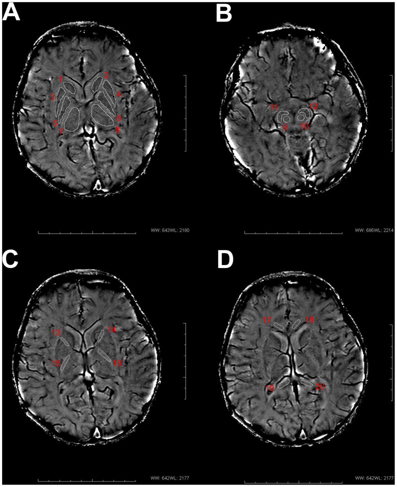
Figure 1. Regions of interest (ROIs) in phase images.
ROIs placement is illustrated on images from an infant brain(postmenstrual age = 90 w)(A) 1, 2 : caudate nucleus; 3, 4: putamen; 5, 6: globus pallidus; 7, 8: thalamus. (B) 9, 10: red nucleus; 11, 12: substantia nigra. (C) 13, 14: anterior limb of the internal capsule; 15, 16: posterior limb of the internal capsule. (D) 17, 18: genu of the corpus callosum; 19, 20: splenium of the corpus callosum.

