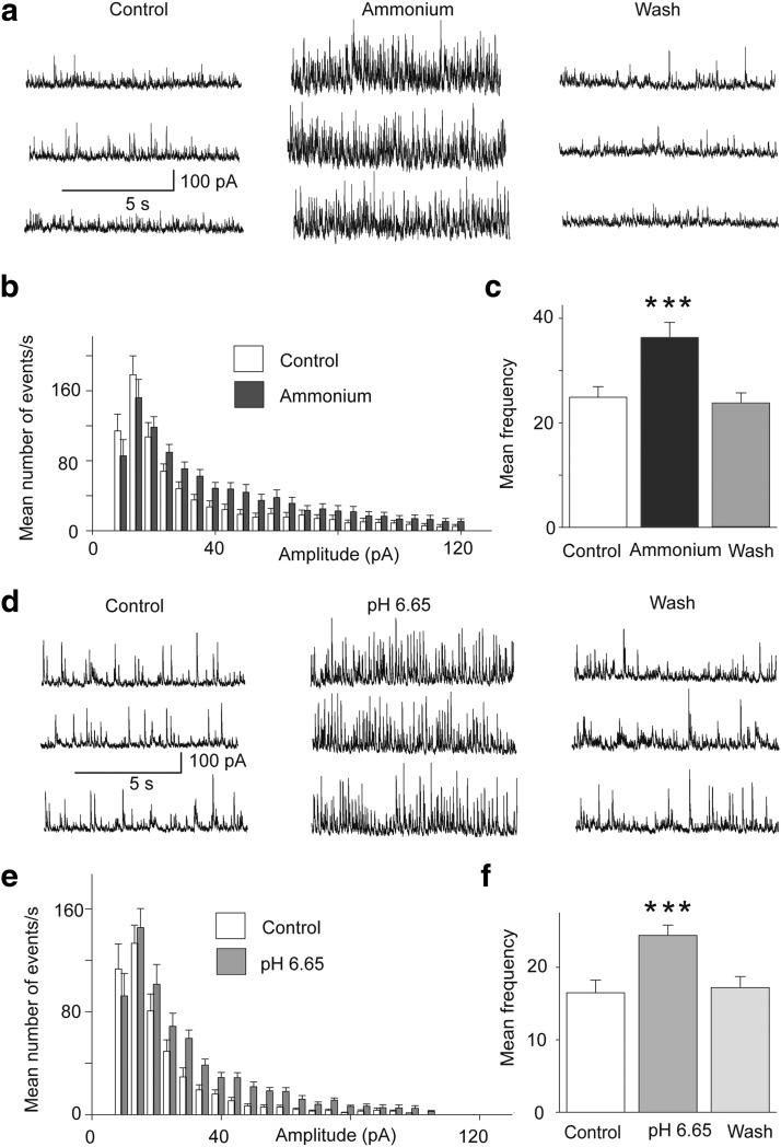
Figure 3.
Activation of ASIC1a increases spontaneous inhibitory activity. Recordings were obtained from BLA principal cells in the presence of CNQX (10 μm), d-AP5 (50 μm), and SCH50911 (10 μm), at Vh = +30 mV. a, sIPSCs before, during, and after bath application of 5 mm ammonium. b, Amplitude-frequency histogram of sIPSCs before and after bath application of 5 mm ammonium (n = 11 neurons from 4 rats); bin width is 5 pA. c, Group data of the frequency of sIPSCs in control medium, in 5 mm ammonium, and after washing out of ammonium (n = 11); ***p < 0.001 when compared with the control. d, sIPSCs before, during, and after perfusion of the slices with acidified ACSF. e, Amplitude-frequency histogram of sIPSCs in control medium and in pH 6.65 (n = 8 neurons from 3 rats); bin width is 5 pA. f, Group data of the frequency of sIPSCs in control medium, in low pH, and after return to control medium (n = 8); ***p < 0.001 when compared with the control.

