Figure 6.
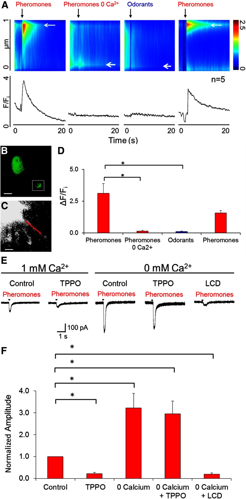
Dependence on extracellular Ca2+ of influx of Ca2+ into the cilia of the TRPM5-GFP+ OSNs and related loose-patch response of TRPM5-GFP+ OSNs to pheromones. A–D, Intraciliary Ca2+ responses of TRPM5-GFP+ OSNs to pheromones require extracellular Ca2+. A, Top, Discrete change in fluorescence (pseudocolor) elicited by pheromones along the proximal 1 μm ciliary segment of the cilium (ordinate) induced by the chemical stimulus as a function of time (abscissa). The color code bar is shown to the right. Bottom, F/Fi as a function of time. B, GFP fluorescence from the TRPM5-GFP+ OSN used to generate the data shown in A. C, Higher-magnification X-Rhod-1 fluorescence image of the dendritic knob and cilia. The red line denotes the segment of the cilium where the line-scan fluorescence recording in C was obtained. D, Bar plot of the peak ΔF/Fi results from five cells. Asterisks denote significant differences in ΔF/Fi (Wilcoxon rank sum test, p < 0.0001, pFDR = 0.03; n = 5). Error bars indicate SEM. E, F, Dependence on extracellular Ca2+ of loose-patch responses to pheromones of TRPM5-GFP+ OSNs. E, Responses to pheromones by a TRPM5-GFP+ OSN in Ringer's solution with 1 and 0 mm Ca2+ Ringer's solution. Under 1 mm Ca2+, it was treated with TPPO, and under 0-Ca2+ it was treated with TPPO and then with l-cis-diltiazem. F, Peaks of the responses to pheromones of TRPM5-GFP+ OSN as in E. Asterisks indicate Wilcoxon rank sum test differences (p < 0.015, pFDR = 0.05; n = 12).

