Figure 5.
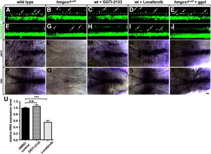
Protein prenylation is required to target dorsally migrating OPCs to the DLF. A–J, Lateral views, with dorsal up, of trunk spinal cords of living 3 dpf larvae. Whereas dorsal OPCs (arrows) cluster near the DLF (dotted line) in wild-type (A), OPCs occupy ectopic position in dorsal spinal cord of a hmgcs1vu57 mutant larva (B), a wild-type larva treated with the geranylgeranyl transferase inhibitor GGTI-2133 (C), and a wild-type larva treated with the farnesyl transferase inhibitor Lonafarnib (D). OPCs occupy normal positions near the DLF in a hmgcs1vu57 mutant larva injected with ggol (E). F–J, Images of larvae carrying the Tg(nkx2.2a:EGFP-CaaX) reporter. In wild-type (F), nascent myelin membrane on dorsal, longitudinal axons is revealed by membrane-tethered EGFP (brackets). In a hmgcs1vu57 mutant larva (G) no membrane localization is evident and EGFP appears cytosolic in OPCs (arrows). Membrane localization of EGFP appears normal in a GGTI-2133 treated larvae (H) but absent in a Lonafarnib treated larva (I). Ggol injection into a hmgcs1vu57 mutant larva rescues membrane localization (J). K–T, Images of hindbrains viewed from dorsal with anterior to the left, of larvae processed for in situ RNA hybridization. Similar to wild-type (K) and by contrast to a hmgcs1vu57 mutant (L), wild-type larvae treated with GGTI-2133 (M) and Lonafarnib (N) express plp1a at high level. Ggol injection also rescues plp1a expression in a hmgcs1vu57 mutant larva (O). mbp expression appears similar to wild-type control in inhibitor-treated larvae (P–S) and is also rescued in a hmgcs1vu57 mutant larva by ggol injection (T). U, Graph showing mbp RNA expression levels in 4 dpf larvae measured by quantitative RT-PCR. mbp expression is not affected by GGTI-2133 but is reduced ∼50% by Lonafarnib (n = 4 biological replicates consisting of 10–15 larvae for each measurement; ***p < 0.0005, unpaired two-tailed Student's t test). Error bars represent SEM. Scale bars, 20 μm.

